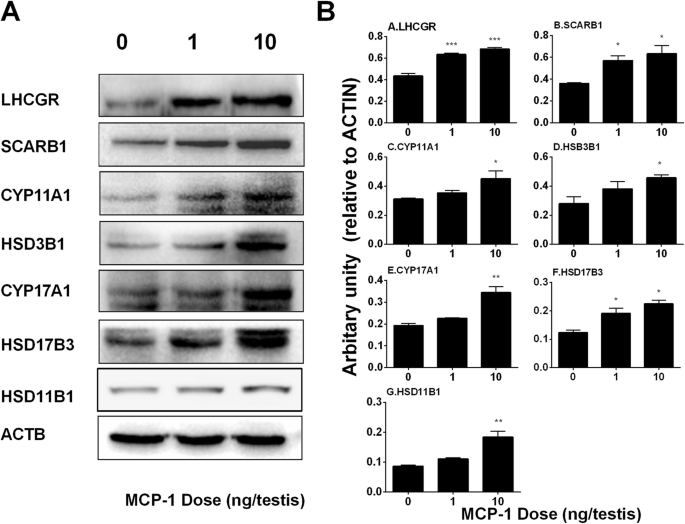
Morphology of Leydig cells in the testes after in vivo MCP-1 treatment.
Por um escritor misterioso
Last updated 14 maio 2024

Monocyte Chemoattractant Protein-1 stimulates the differentiation of rat stem and progenitor Leydig cells during regeneration, BMC Developmental Biology
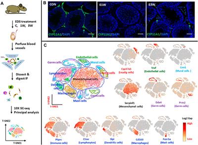
Frontiers Identification of Rat Testicular Leydig Precursor Cells by Single-Cell-RNA-Sequence Analysis

Morphology of Leydig cells in the testes after in vivo PTHrP
Frontiers Insights into the Development of the Adult Leydig Cell Lineage from Stem Leydig Cells

Fluoride-Induced Autophagy via the Regulation of Phosphorylation of Mammalian Targets of Rapamycin in Mice Leydig Cells
Molecules, Free Full-Text
Frontiers Cytokines in Male Fertility and Reproductive Pathologies: Immunoregulation and Beyond
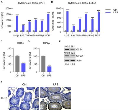
Frontiers OCT4 Represses Inflammation and Cell Injury During Orchitis by Regulating CIP2A Expression
Frontiers Mumps Orchitis: Clinical Aspects and Mechanisms
Sertoli cell ablation in adulthood induces apoptotic loss of Leydig

SARS-CoV-2 infection leads to sustained testicular injury and functional impairments in K18 hACE2 mice

Morphology of Leydig cells in the testes after in vivo MCP-1 treatment.

Morphology of Leydig cells in the testes after in vivo MCP-1 treatment.

Antioxidants, Free Full-Text
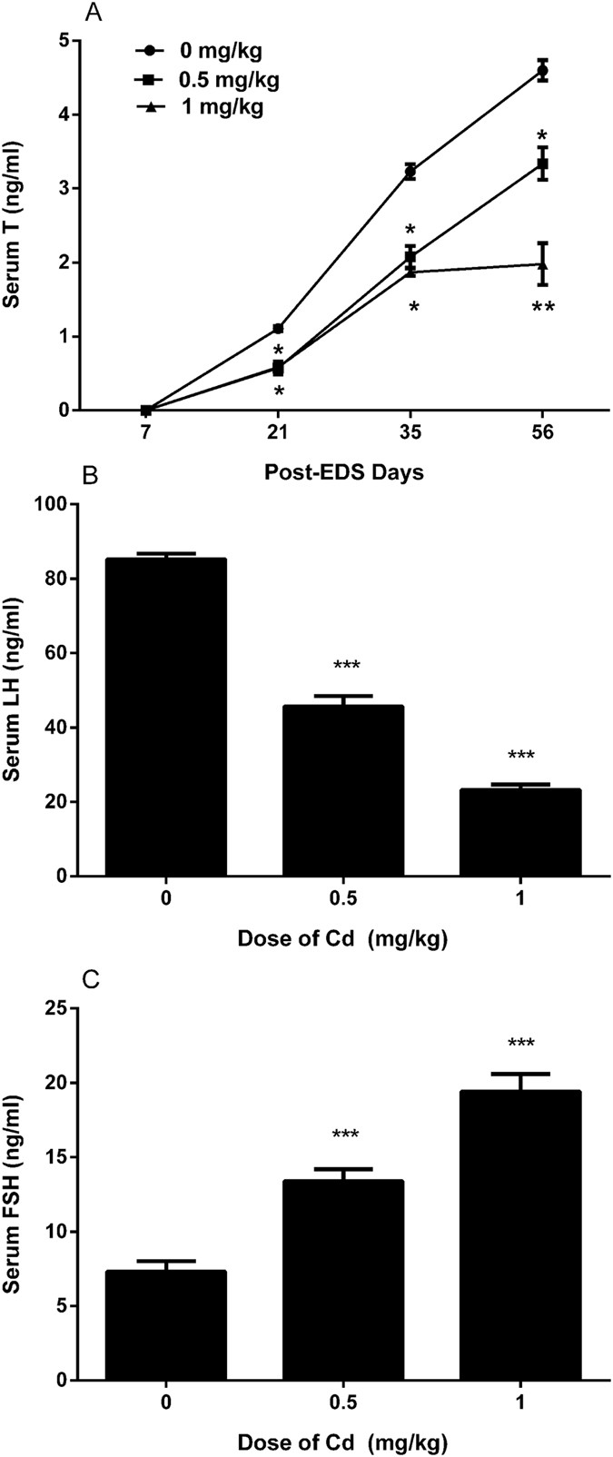
A brief exposure to cadmium impairs Leydig cell regeneration in the adult rat testis
Recomendado para você
-
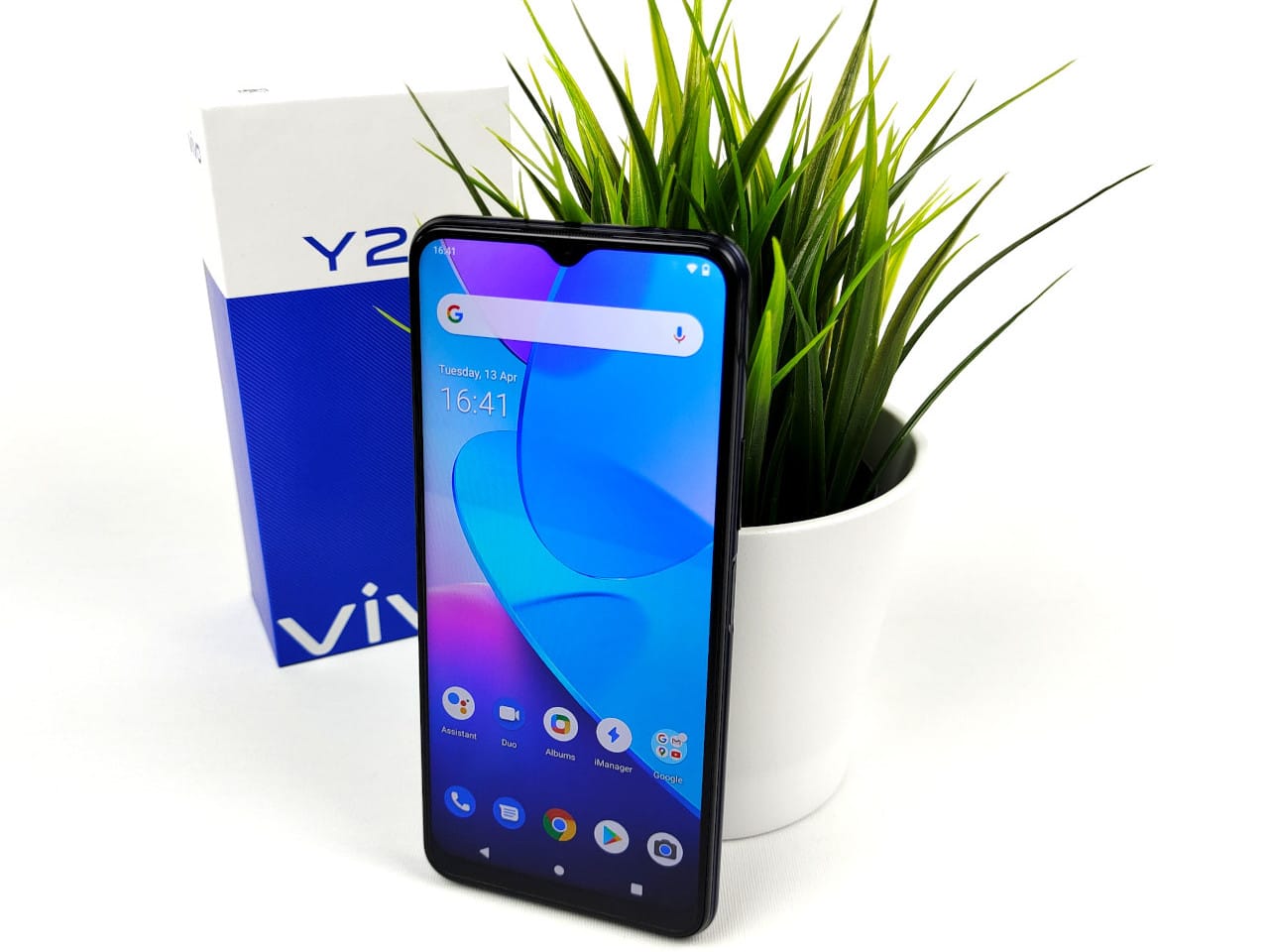 vivo Y20s no teste - o smartphone vivo mais barato14 maio 2024
 -
 Teste Palográfico: da técnica à prática - Grupo Educativa14 maio 2024
 -
 solteiro triste mulher reclamando segurando uma gravidez teste sentado em uma sofá dentro a vivo quarto às lar. depressivo Preto menina segurando negativo gravidez teste. 20419033 Foto de stock no Vecteezy14 maio 2024
 -
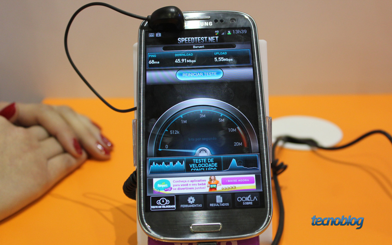 Vivo relança TV via fibra óptica e começa testes com 4G – Tecnoblog14 maio 2024
 -
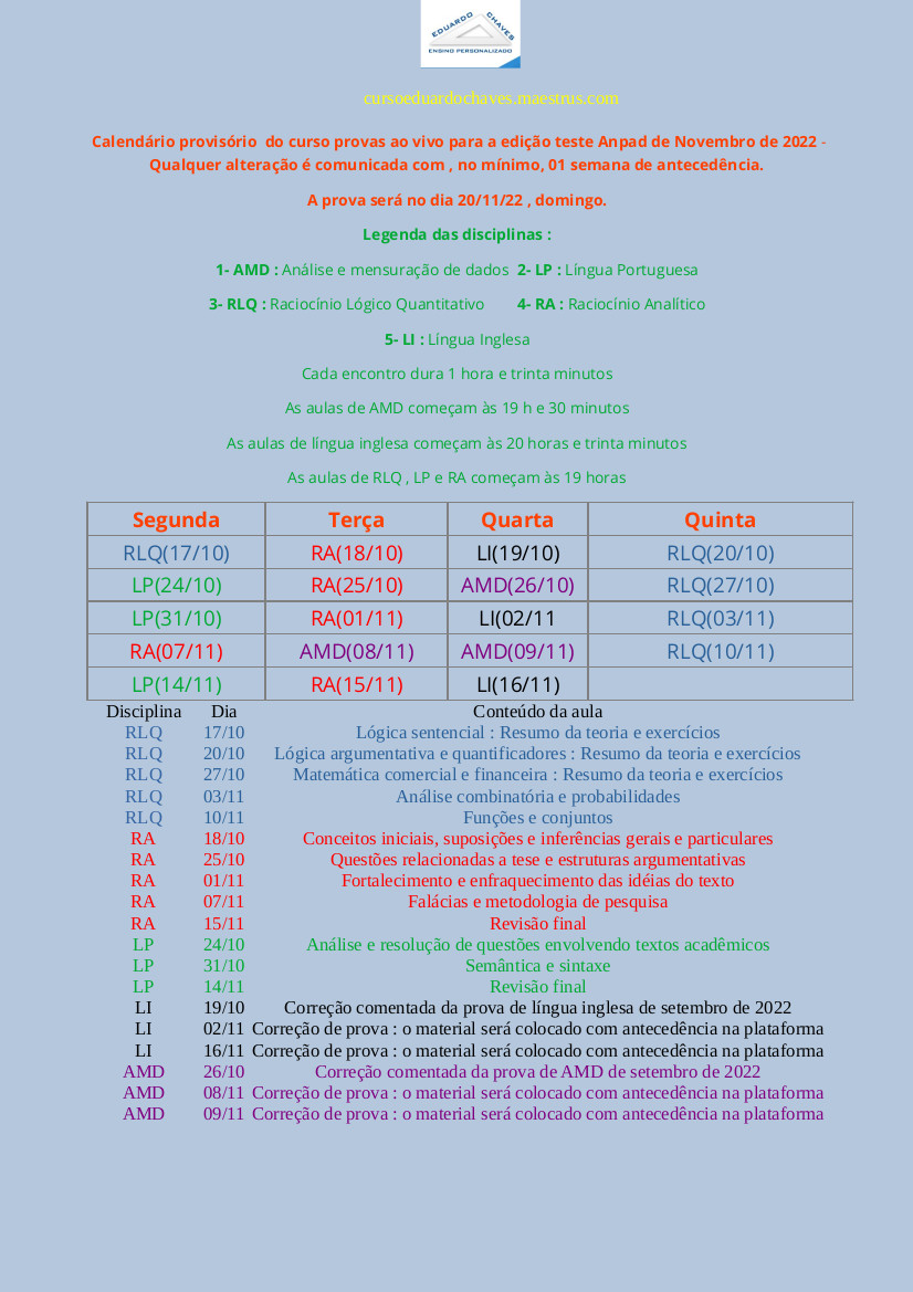 Teste Anpad 2022 - 2023 - Curso de provas ao vivo para a edição de novembro de 202214 maio 2024
 -
 The Division Resurgence: O próximo teste ao vivo começará em 8/1214 maio 2024
 -
 Botão webinar ao vivo. ícone de cor azul para curso online, educação a distância, vídeo-aula, conferência em grupo na internet, teste de treinamento. webinar ao vivo com microfone, ícones de transmissão14 maio 2024
 -
 TESTE GRÁTIS 24 HORAS internet ilimitada da VIVO14 maio 2024
 -
 Multímetro digital Testador de caneta inteligente Caneta de capacitância de tensão de autoranging Medidor de teste elétrico Diodo-Continuidade Medidor ao vivo Testador de circuito-Sonda Ferramenta elé14 maio 2024
 -
 Mini Teste Ao Vivo 1550nm 20db Da Fibra Ótica Do Teste Ativo De14 maio 2024
você pode gostar
-
 ORAÇÃO DO DIA-27 DE SETEMBRO @BispoBrunoLeonardo : News Informa .14 maio 2024
 -
 World Cup 2022 quarter-final fixtures: Qualified teams & kick off14 maio 2024
 -
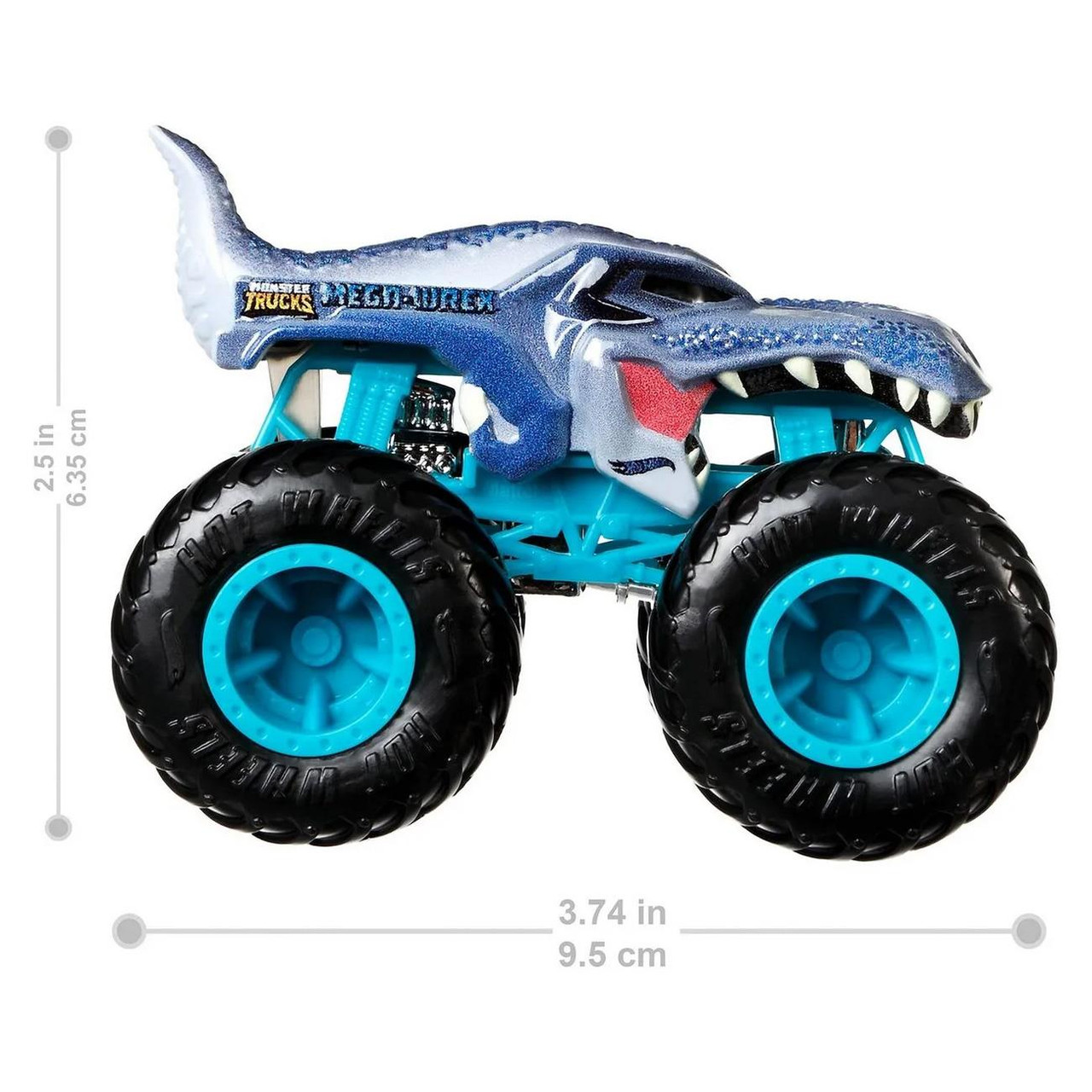 Hot Wheels Monster Trucks Demolition Doubles MOTOSAURUS vs. MEGA WREX 1:64 Scale Vehicle 2-Pack14 maio 2024
 -
 POKEMON CARD - KARTANA GX FULL ART 106/111- CRIMSON INVASION NM14 maio 2024
 -
Top 5 Áreas Masculinas Mais Procuradas para Remoção a Laser14 maio 2024
-
 Você sabe sobre a América do Sul e seus países e cidades?14 maio 2024
 -
 Topo De Bolo Maquiagem 3 - Pronto No Palito14 maio 2024
 -
 Dream World — Among Us Cookies14 maio 2024
 -
Dragon Ball Clássico Pura Nostalgia10014 maio 2024
-
 Fogelman Focus Magazine 2023 by University of Memphis - Issuu14 maio 2024
