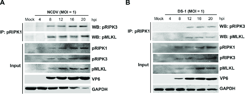
RVA-induced necrosome formation consisting of RIPK1, RIPK3, and MLKL
Por um escritor misterioso
Last updated 27 junho 2024

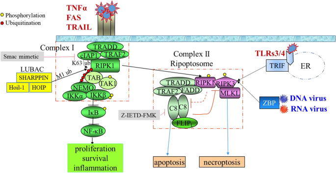
Overview of necroptotic signaling pathway. Necroptosis can be induced
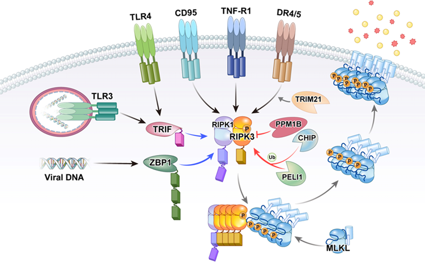
Roles of RIPK3 in necroptosis, cell signaling, and disease
RIPK3 signaling and its role in the pathogenesis of cancers

From (Tool)Bench to Bedside: The Potential of Necroptosis Inhibitors

From (Tool)Bench to Bedside: The Potential of Necroptosis Inhibitors

Opposite Effects of Apoptotic and Necroptotic Cellular Pathways on Rotavirus Replication

RIPK3 - an overview ScienceDirect Topics

MLKL in cancer: more than a necroptosis regulator. - Abstract - Europe PMC
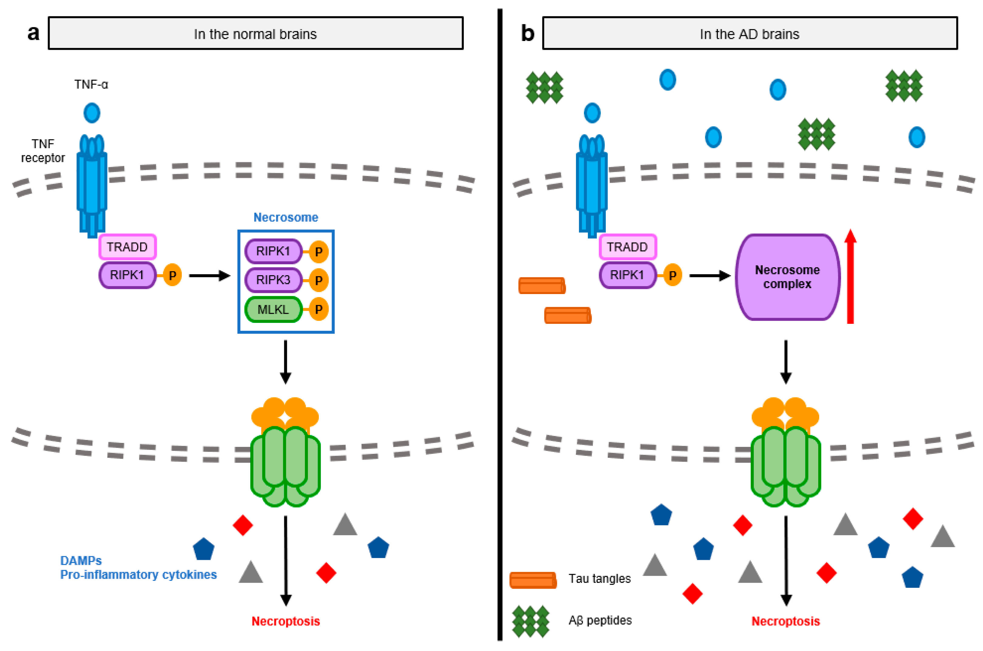
IJMS, Free Full-Text
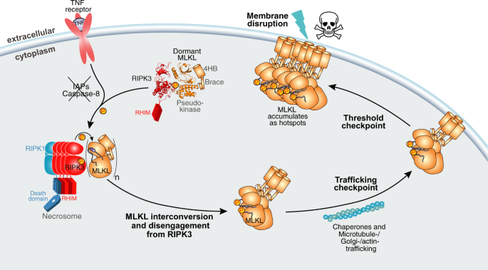
Human RIPK3 maintains MLKL in an inactive conformation prior to cell death by necroptosis
Recomendado para você
-
 Números 14:28 RVA - Diles: Vivo yo, dice Jehová, que según habéis27 junho 2024
 -
 Illustration of viscosity property using rapid visco analyzer (RVA27 junho 2024
 -
 detector de movimento Sistema de Câmera de Segurança – Empresa de27 junho 2024
 -
Anupam Sharma posted on LinkedIn27 junho 2024
-
 CELEBRAÇÃO AO VIVO, Cidade Viva Brasília27 junho 2024
 -
Synthesis and Biological Evaluation of Enantiomerically Pure (R27 junho 2024
-
US-Mexico the Nations League Final we expected, but not how we27 junho 2024
-
 The structure, properties and potential probiotic properties of starch-pectin blend: A review - ScienceDirect27 junho 2024
 -
 Rádio Venus - Portal RVA27 junho 2024
 -
 Pesquisa Nupes/Unisc confirma RVA AM e Venus FM como líderes de27 junho 2024


você pode gostar
-
lh3.googleusercontent.com/t_ZZoH_oSUOGyMcM8dGOxZJ927 junho 2024
-
Lemmy on X: parry this filthy casual [farfetch'd (kanto and galar) redesigns! rts appreciated] / X27 junho 2024
-
 Star Platinum Full Muscle Costume Jojo Kujo Jotaro Stand Cosplay Suit - Gloden Line version full body muscle stuffed27 junho 2024
 -
Quiz Infantil – Apps no Google Play27 junho 2024
-
 FNAF SB Ruin - MXES The Entity Art Board Print for Sale by27 junho 2024
 -
 Death Note's Kira Cameo in Death Parade?27 junho 2024
 -
 Dive into the Past! The Fascinating History of Restaurant Booths!27 junho 2024
 -
 Dont Pay Tax Stock Photos - Free & Royalty-Free Stock Photos from Dreamstime27 junho 2024
 -
 Ryang-ha Song, Koroshi Ai Wiki27 junho 2024
 -
 Free Vector, Fire flame burn flare torch hell fiery icons set isolated vector illustration27 junho 2024

![Lemmy on X: parry this filthy casual [farfetch'd (kanto and galar) redesigns! rts appreciated] / X](https://pbs.twimg.com/media/Ey0IyNJWgAE1fxE?format=jpg&name=4096x4096)